Compare costs
Average cost of treatment in key countries
Include indicative costs for treatment, travel, insurance and accommodation
Get a quote
1. Complete the enquiry form
2. Select countries of interest
3. Providers respond directly
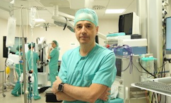
Based at two leading hospitals in Madrid, Dr Ricardo Diez Valle Neurosurgery offers expert diagnosis, management and treatment for conditions affecting the brain from an internationally recognised team. Equipped with the latest technology and adopting the most recent advances in the field, including minimally invasive cranial surgery and cranial microsurgery, they provide individualised treatment for conditions such as malignant and benign brain tumours, cavernomas and trigeminal neuralgia.
 The neurosurgery team of Dr Diez Valle aims to provide its patients with the most up-to-date and precise neurosurgical treatment. The best national and international training and the most sophisticated technology are put at the service of each patient's treatment, allowing for the planning and execution of the individualised surgery with maximum effectiveness and minimal risk.
In modern neurosurgery, technological developments and biological advances converge to offer continuous improvements, often small but occasionally significant. The outcome that can be achieved in neurosurgical interventions varies widely and depends on many details that need to be carefully managed.
The team of Dr Diez Valle is internationally recognised for its ability to apply scientific advances (technological and biomedical) rapidly to the treatment of its patients. He is a world pioneer in the application of fluorescence-guided surgery with fluorescence and intraoperative MRI, minimally invasive cranial surgery, immunotherapy and the use of oncolytic viruses, as reflected in his CV and publications.
The experience gained over more than 25 years allows Dr Diez Valle and his team to perform microsurgery that is minimally invasive without sacrificing effectiveness. The Keyhole technique offers a smaller scar and preservation of healthy tissue, resulting in faster recovery with minimal discomfort and complications. This concept has been successfully applied and refined throughout his professional career at the Ramon y Cajal University Hospital, the MD Anderson Center and the University of Navarra Clinic, with multiple international collaborations.
The team has accumulated experience in more than 2000 microsurgeries for cranial tumours and is currently employing this technique routinely, using the most advanced visualisation platform on the market, which combines robotic microscope, exoscope and endoscope in the same system.
In neurosurgery, especially in brain surgery, each patient is different, with a unique problem, and there are no two identical situations. Therefore, the Center’s sophisticated equipment is essential for studying, planning and executing each surgery. High Field, 3T MRI combined with tractography allows a detailed map of the brain around the lesion.
But above all that, it is essential to combine technological considerations with a meticulous assessment of the human aspects of each patient, listening to and respecting their preferences and expectations. Only then is it possible for each patient to receive the alternatives and choose their best option.
In each case, Dr Diez Valle and his team seek personalised therapy, i.e. the most suitable for each patient's situation.
Dr Diez Valle has designed and been principal investigator of clinical trials with immunotherapy: dendritic cell vaccines, injection of oncolytic viruses, and trials with immunomodulators.
 State-of-the-art hospitals
Dr Diez Valle performs his surgeries at two leading hospitals in Madrid. Both have state-of-the-art operating theatres equipped for complex brain surgery.
Low-grade gliomas are a group of tumours characterised by slow growth. Despite their slow growth, they still pose a significant problem for the patient as they gradually damage the brain and can result in neurological damage, disability and even death if their growth is not stopped. They most commonly appear in young adults, between 20 and 50 years old and their origin is unknown; they are not associated with any known risk factors, nor associated with known genetic characteristics.
Symptoms of a low-grade glioma can appear insidiously over time or suddenly. Symptoms include progressive headaches, loss of strength or sensation, language alteration, among others. Often the first symptom is an epileptic seizure. These tumours are identified using MRI and CT, however analysing the tissue is crucial to determine the exact subtype of the tumour to pin-point the correct course of treatment.
Treatment plans for diagnosed low-grade glioma must take into account all aspects of the tumour’s exact biology, location and size, as well as the patient’s clinical status, age and expectations. Based on the most recent research, Dr Diez Valle always recommends surgical removal rather than conservative treatment and monitoring. Each surgery is carefully planned to achieve maximal surgery with minimal risk. Techniques used include minimally invasive microsurgery which allows a patient to return home within 5 days and interoperative high-field MRI in complex cases. Following tumour analysis, a treatment continuation plan is established together with medical and radiation oncology, if required.
Glioblastomas (GBM) are an aggressive, fast growing brain tumour.There are no known risk factors for developing these tumours. Symptoms can appear within a few days or weeks and include headaches, retroocular pressure, nausea and vomiting, seizures, loss of strength or sensation in the limbs, language impairment and visual and behavioural disturbances.
Diagnosis of GBM can be made on imaging studies, particularly MRI and high-field MRI. However, analysis of the tissue is necessary to confirm and determine the exact type of tumour.
Due to their aggressive nature, overall prognosis for a GBM is poor, although patients can benefit from treatments that improve overall survival and quality of life.As part of multidisciplinary care, neurosurgery for GBM has been shown to be beneficial both for symptom improvement and patient survival and should be the first step in the treatment process.
Dr Diez Valle is experienced in the technically challenging and advanced surgical techniques necessary to perform complete resection of glioblastomas, including surgical neuronavigation, intraoperative MRI, intraoperative ultrasound and fluorescence-guided surgery.
Meningiomas are the most common benign cranial tumours. In most cases the cause of their origin is unknown, however in a very few cases it is a genetic condition. They are slow growing and may not produce any symptoms for years. Eventually symptoms may appear, the most general being a headache.
Meningiomas are usually diagnosed by CT or MRI. Once diagnosed, there are three treatment paths: monitoring only with periodic MRI scans; microsurgery to remove the tumour; or radiosurgery.
Surgery can be curative and is indicated for young patients, if the tumour is large or if the tumour is causing discomfort. Minimally invasive microsurgery for meningioma results in faster recovery, minimises complications and reduces discomfort. Patients can return home 2-5 day after surgery.
Radiosurgery is an alternative treatment in which a targeted dose of radiation is delivered to the tumour in a single session. The advantage is that there is no wound or scar, and the risks of injury are very low, although not zero. The disadvantages are that, although the tumour usually stops growing, it doesn’t disappear and will need continued monitoring. Also, the tumour is not analysed, so there is a small risk that it may be another type of tumour. Radiosurgery results can be excellent especially when performed with the most sophisticated technology, such as GammaKnife. It may be the best option for small tumours in difficult locations or in patients at very high risk. The technique can also be used in conjunction with surgery for large tumours in complicated locations.
Dr Diez Valle offers personalised treatment options for patients with meningioma and work with the highly experienced Gamma Knife Unit at Ruber International Hospital to offer the best of both options, or the combination.
Vestibular schwannomas are slow growing, benign tumours that originate from Schwann cells which form the covering of nerves. They can arise spontaneously, although occasionally they may be associated with genetic alterations, being very common in the condition, neurofibromatosis.
Symptoms of schwannomas usually appear subtly and progress slowly in relation to the compression of the nerve from which they originate and the surrounding structures. The most frequent location is the vestibular nerve. Due to its proximity, the auditory nerve will also be affected, and it is for this reason that these tumours are called acoustic neuromas. Symptoms include dizziness, hearing loss, tinnitus, instability and headache. Other schwannomas are less common, but can originate from the trigeminal nerve, facial nerve, glossopharyngeal nerve and nerve roots exiting the spinal cord.
Diagnosis of a vestibular schwannoma is made with a cranial CT or MRI, sometimes with biopsy. Treatment is tailored to the individual; options include surveillance only with periodic MRI scans, microsurgery, radiosurgery or a combination of both.
A brain metastasis is the growth of a malignant tumour in the brain originating from another part of the body. It can occur with any type of cancer, but the most common sources are lung cancer, breast cancer and melanoma. Brain metastases typically grow rapidly often causing significant brain swelling (edema) and symptoms such as headaches, seizure and nausea. Failure to address them properly can lead to rapid symptom progression and potentially death.
Brain metastases are usually diagnosed by CT or MRI. Treatment depends on a number of factors including the type and stage of the primary tumour, the number, location and size of the brain lesions and the patient’s clinical status.
A comprehensive understanding of these factors is crucial for developing an individualised treatment plan for each patient, typically involving multiple specialists. Treatment options may include medication, surgery or radiotherapy.
Trigeminal neuralgia is an irritation of the trigeminal nerve that produces pain in the region of the face innervated by this nerve, typically localised on one side of the face. Often, there is no specific lesion or disease causing the trigeminal nerve irritation – this is termed idiopathic neuralgia. Most of the idiopathic cases are believed to originate from nerve compression caused by a normal, non-pathological vascular structure. However, there are other possible causes leading to these symptoms and a good diagnosis is essential to apply optimal treatment.
Once the diagnosis of trigeminal neuralgia is confirmed, initial treatment always involves medication. Second line options are microvascular decompression, percutaneous ablative procedures (thermocoagulation or balloon compression) or radiosurgery.
Microvascular decompression surgery is the most effective long-term treatment, although it carries the highest short-term risk. More than 90% of patients respond well, resuming normal activities within 3-4 weeks and gradually tapering off pain medication. Pain recurrence occurs in around 10% of patients at 5 years and nearly 20% at 10 years.
Percutaneous ablative treatments pain relief in nearly 90% of cases and are less invasive compared to open surgery. However, as the recurrence rate is approximately 50% at 10 years and nerve damage may occur, the technique is best suited to elderly patients unsuitable for major surgery.
Radiosurgery with Gamma Knife also produces partial damage to the trigeminal nerve and pain cessation. It is a low-risk procedure, but its effects may take time to manifest and the long-term outcomes are less well-known. It is typically considered when decompression and percutaneous procedures have failed or cannot be performed.
Intraventricular tumours grow inside the inner cavities of the brain, called cerebral ventricles, where cerebrospinal fluid is produced and circulates. They are rare, accounting for less than 1% of intracranial lesions. The causes are not well understood; there are some genetic conditions that tend to develop tumours in the ventricles, but they represent a very small percentage of cases.
Symptoms of intraventricular tumours can vary widely. If they obstruct the circulation of cerebrospinal fluid, they can produce symptoms of hydrocephalus, including headaches, nausea, vomiting, altered level of consciousness, instability, and visual disturbances. If they do not obstruct circulation, they can grow very large until they cause symptoms due to compression of adjacent structures, which can include motor or sensory disturbances, or memory impairment.
Most of these tumours are benign and slow growing. When they produce symptoms and the patient seeks medical attention, a CT scan or brain MRI is usually requested. Brain MRI is important not only for tumour diagnosis but also for surgical planning.
Treatment usually involves surgery because surgery allows not only the removal of the tumour, but also precise diagnosis and resolution of hydrocephalus if present.
Surgery for ventricular tumours is challenging for surgeons due to their location in hard-to-reach areas surrounded by vital structures. It is crucial to choose the most appropriate approach for each case, and there are many variations. The skill in using a microscope and the ability to perform minimally invasive approaches are fundamental. If done correctly, the amount of brain tissue damaged to remove the tumour is minimal.
Often, an external ventricular drain needs to be placed just before, during, or after surgery. In some cases, a shunt may need to be implanted to drain the fluid permanently. Recovery may be more challenging than after surgery for brain tumours in other places because it takes some additional time for the cerebrospinal fluid circulation to fully restore. It is common to experience headaches, discomfort, and even some fever in the first few days. However, in cases of benign tumours, recovery can be complete.
Cavernomas are vascular lesions, not tumours, but abnormalities of blood vessels. While it is most common for them to occur in isolation, with any known cause of family history, up to 20% of patients have a hereditary familial form in which multiple lesions appear. This is known as familial cavernomatosis and can be confirmed by genetic testing. They may also occur after brain radiotherapy treatment.
In many cases cavernomas are incidental findings and remain completely asymptomatic. Cavernomas can bleed and grow causing symptoms such as epilepsy, headache, nausea, weakness or numbness on one side of the body, language impairments and disturbances of vision and balance.
When cavernomas bleed, MRI is the definitive test for studying the relationship of the lesion with the normal brain, which is crucial for therapeutic decision making.
Surgical resection is curative for cavernomas. Depending on their location and the problems they have caused, surgery may be recommended or simply surveillance advised. Anti-epileptic drug treatment may be an option to prevent seizures if they are well controlled and surgery is not desired.
It is recommended to operate on lesions that cause symptoms, those that have bled and those that are surgically accessible. Cavernomas are particularly suitable for minimally invasive surgery. The risks and burden of this surgery are low, so it may be better option than living with the lesion and its long-term risk, particularly in young people.
| Liability insurance |
No |
