Dr Manchon Diagnostic Imaging Centre
Imaging centre in Barcelona, Spain
PROFILE
Dr Manchon Diagnostic Imaging Centre in Barcelona, Spain, offers excellent CT scans, MRI scans and diagnostic imaging at very competitive prices.
Although MRI and CT scans are the key aspects of their activity they also provide x-ray examinations, mammography, medical ultrasonography (sonography), bone density measurements. As well as searching for current disorders or their causes they provide health screening for general prevention and early detection.
MRI scans, CT scans, mammography in Barcelona
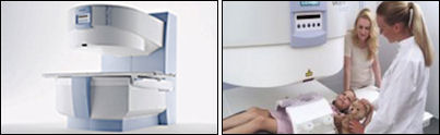
Magnetic Resonance Imaging (MRI scan)
In a magnetic field, reflected waves of radiowaves applied to your body will be received and transformed to very precise images, particularly of the soft tissues (whereas a CT-scan provides detailed information about the structure of bones). The examination usually takes 20 to 30 minutes and you will be closely monitored all the time. To obtain additional information, a contrast agent may sometimes be useful.
The imaging centre in Barcelona, has non claustrophobic magnetic resonance technology. This technology provides maximum comfort for claustrophobic and anxious patients as well as children. Further advantages of this technology:
- Quiet scans
- Easier access
- Comfortable positioning of the patient
- Open on three sides for a panoramic 270° view
Computed Tomography (CT scan)
CT combines the use of x-rays with latest computer technology to create cross-sectional images of your body. The examination lasts from 5-15 minutes, depending on the body region examined.
After the examination, computerised reconstructions can create sectional images in other planes (e.g. vertical). Sometimes, intravenous contrast(dye) is used to highlight blood vessels and to enhance the structure of organs like the brain, spine, liver, and kidney.
Mammography
Cancer of the breast is very common. In Mammography the breast is examined with a specific x-ray unit, that applies very soft x-rays. Different kinds of tissue can be displayed with high contrast, especially small calcifications which are often early signs of breast cancer.
X-ray examination
X-ray, or radiography, is the easiest way for a physician to assess broken bones; however, x-ray is used for more than bones and joints. An x-ray image is produced, when a small amount of x-rays passes through your body and strikes a sensitive film on the other side of the body, which will thus be darkened. The tissues of your body absorb the rays to a less extent than bones, while the air containing lungs let most of the rays pass unchanged. The film will show a shadow-like image of the contents of your body in a grey scale (with bones almost white, lung almost black).
Medical ultrasonography (sonography)
This imaging technique is used to obtain a first insight into muscles and internal organs, their size, structures and any pathological lesions, it can be very useful in the hands of an experienced examiner, particularly for slim patients. In case of lesions detected, sometimes secondary examinations become advisable, like follow up examinations or crossectional studies. Sonographers typically use a hand-held probe (called a transducer) that is placed directly on and moved over the patient.
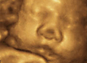 3D-4D Ultrasound
RealTime 4D imaging is the continuous, three dimensional scanning of an object with simultaneous visualization of the A, B and C planes. This revolutionary quad-beam technology, obtaining 3D images in RealTime up to 25 volumes per second, goes beyond the boundaries of traditional ultrasound and opens the 4th dimension of time to the medical world.
This technology gives the radiologist additional information for a more accurate diagnosis and highest quality care.
Obstetrics patients have an option to have images and video on a CD-Rom and 3D photos, you can see incredible fetal images (3D ultrasound) and real-time movements (4D ultrasound).
Bone densitometry
Bone density measurements at the Dr Manchon imaging center are carried out using the latest densitometer - the dual photon energy (DXA) Discovery QDR densitometer. It has been scientifically proven that dual photon energy technology is the best method for bone density measurement. This machine takes spine as well as femur density measurements.
Legal
| Liability insurance: | No |
CONTACT US
|
Avinguda del Tibidabo 9 Barcelona Catalonia 8022 Main phone+34934450600 Fax+34932530780 |
|