Radiologie Ettlingen
Imaging centre in Ettlingen, Germany
PROFILE
Radiologie Ettlingen, a diagnostic imaging centre in Ettlingen, Germany offers excellent CT scans, MRI scans and diagnostic imaging at very competitive prices.
Although MRI and CT scans are the key aspects of their activity they also provide x-ray examinations, mammography, medical ultrasonography (sonography), bone density measurements and nuclear medicine (scintigraphy). As well as searching for current disorders or their causes they provide health screening for general prevention and early detection.
Examinations offered by the Ettlingen Diagnostic Imaging Centre
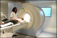
Magnetic resonance imaging (MRI scan)
Radiologie Ettlingen uses a high performance Siemens Avanto MRI System with Syngo, EPI, and the complete Siemens Advanced Imaging Equipment including DESS and CISS sequences. The system was installed 9/2010 representing the latest technology.
In a magnetic field, reflected waves of radiowaves applied to your body will be received and transformed to very precise images, particularly of the soft tissues (whereas a CT-scan provides detailed information about the structure of bones). The examination usually takes 20 to 30 minutes and you will be closely monitored all the time. To obtain additional information, a contrast agent may sometimes be useful.
CT (Multi Slice Computed Tomography) scan
MSCT combines the use of x-rays with sophisticated computer technology to create cross-sectional images of your body. A new Siemens Emotion MSCT system is used, also installed 9/2010. The examination lasts from 5-15 minutes, depending on the body region examined.
After the examination, computerised reconstructions can create sectional images in other planes (e.g. vertical). Sometimes, intravenous contrast(dye) is used to highlight blood vessels and to enhance the structure of organs like the brain, spine, liver, and kidney.
Mammography
Cancer of the breast is very common. In Mammography the breast is examined with a specific x-ray unit, that applies very soft x-rays. Different kinds of tissue can be displayed with high contrast, especially small calcifications which are often early signs of breast cancer.
X-ray examination
X-ray, or radiography, is the easiest way for a physician to assess broken bones; however, x-ray is used for more than bones and joints. An x-ray image is produced, when a small amount of x-rays passes through your body and strikes a sensitive film on the other side of the body, which will thus be darkened. The tissues of your body absorb the rays to a less extent than bones, while the air containing lungs let most of the rays pass unchanged. The film will show a shadow-like image of the contents of your body in a grey scale (with bones almost white, lung almost black).
 Medical ultrasonography (sonography)
This imaging technique is used to obtain a first insight into muscles and internal organs, their size, structures and any pathological lesions, it can be very useful in the hands of an experienced examiner, particularly for slim patients. In case of lesions detected, sometimes secondary examinations become advisable, like follow up examinations or crossectional studies. Sonographers typically use a hand-held probe (called a transducer) that is placed directly on and moved over the patient.
Nuclear medicine (Scintigraphy)
For scintigraphic examination, a small amount of radioactive substance is injected into an vein. The distribution in your body can be followed with a Gamma Camera and a computer. The method is very sensitive for changes in bone metabolism and is useful for searching inflammatory or malignant focuses in follow up examinations after cancer disease. Nodal changes in the thyroid can be evaluated, whether they produce more thyroid hormone than normal tissue or less decisive for therapy. In case of suspected embolic lung disease, clogged vessels in the lung can be detected sensitively by scintigraphy, because the substance will not reach all parts of the lung.
Bone densitometry
Bone density measurements at Radiologie Ettlingen are carried out using the latest densitometer - the dual photon energy (DXA) Discovery QDR densitometer. It has been scientifically proven that dual photon energy technology is the best method for bone density measurement. This machine takes spine as well as femur density measurements.
Competitive costs
Costs include flight and local accomodation, if required. Prices charged depend on the part of the body which is examined. The following are indicative prices for MRI examinations:
- Knee or another joint - £250
- Both knees - £400
- Lower, middle, or upper part of the spine - £250
- Whole spine (three parts) - £500
- Head - £300
- Upper Abdomen or Pelvis - £300
- Intravenous contrast media (if required) - £100
Discounts are given if you take several examinations or for groups who undertake examinations on the same day.
Legal
| Liability insurance: | No |
CONTACT US
|
Wilhelmstrasse 1 Ettlingen 76275 Main phone+497243531510 Fax+49724313845 |
|